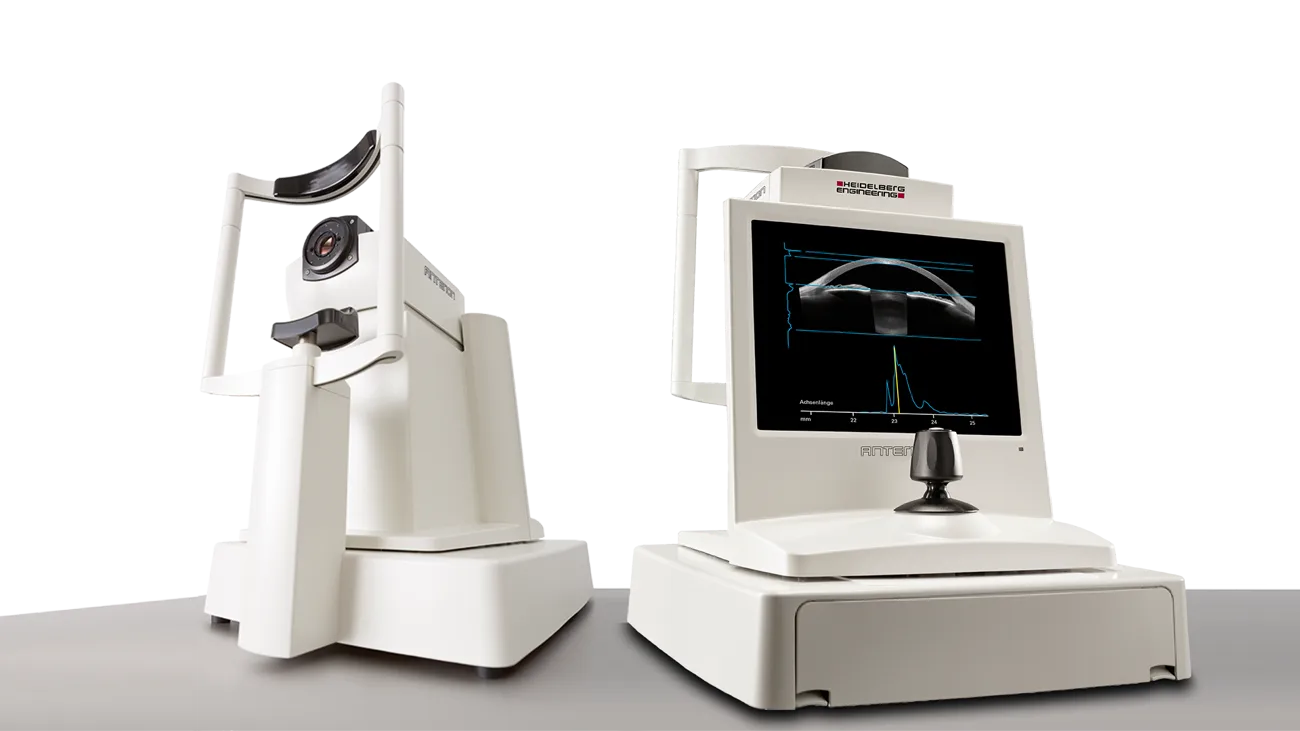
The ANTERION system is an advanced anterior segment imaging platform that uses swept-source OCT technology to deliver high-resolution, cross-sectional images of the eye. It plays a crucial role in evaluating the cornea, anterior chamber, and lens, making it highly valuable for cataract, refractive, and glaucoma assessments.
With its ability to provide detailed measurements and comprehensive analysis in a single scan, ANTERION enhances diagnostic accuracy and treatment planning. Its non-invasive and patient-friendly approach ensures comfort while enabling clinicians to make informed decisions with confidence.
By incorporating technologies like ANTERION, Abate Eye Hospital continues to ensure accurate diagnosis, personalized treatment, and superior eye care outcomes.
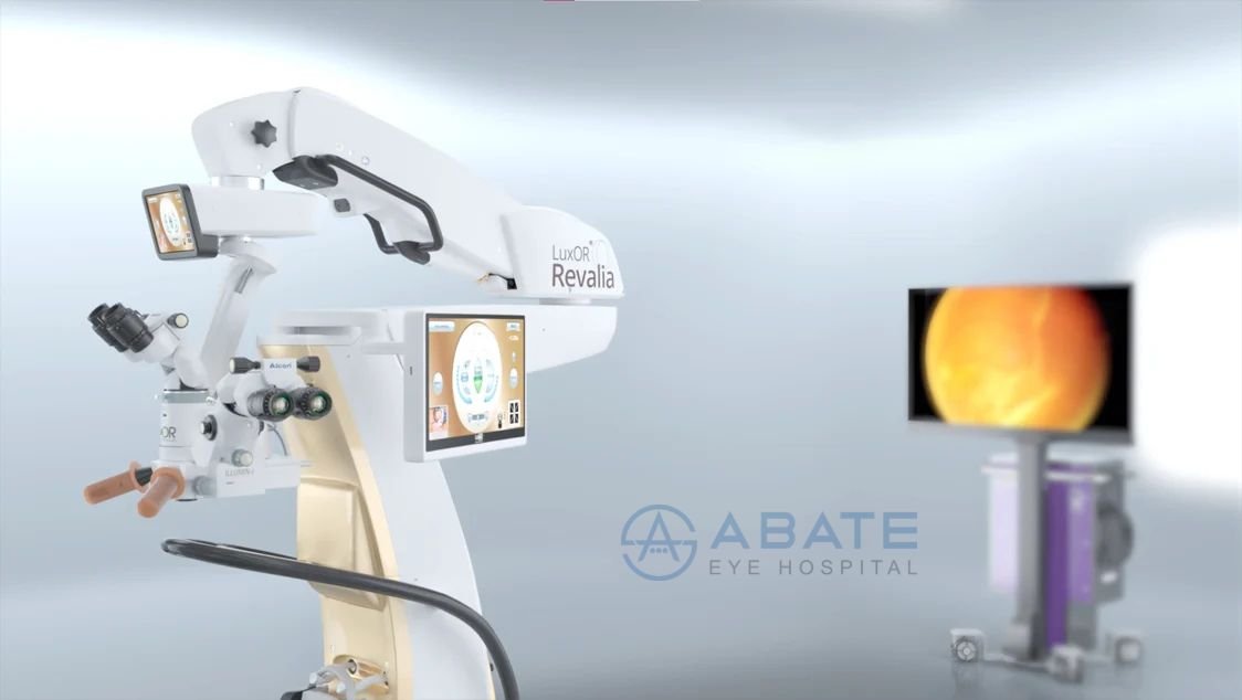
At Abate Eye Hospital, advanced technology plays a vital role in delivering precise and effective eye care. One of the latest innovations available is the ALCON Revalia, designed to enhance surgical accuracy and patient safety.
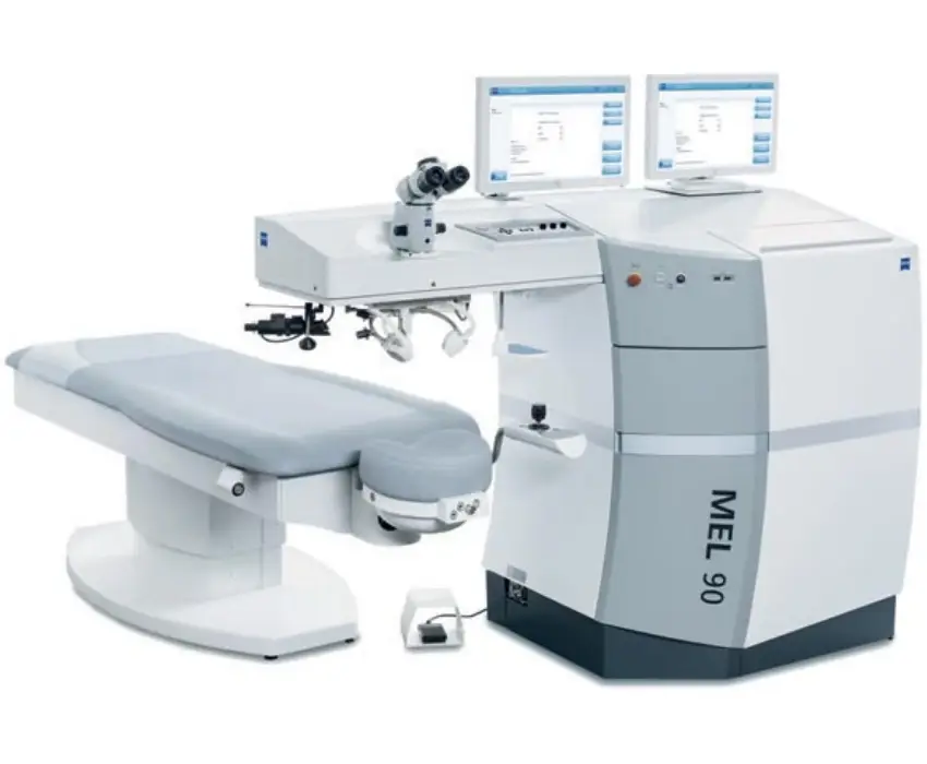
The best equipment is a key component of optimal results. Our hospitals are equipped with the following industry-leading technology:
The best equipment is a key component of optimal results. Our hospitals are equipped with the following industry-leading technology:
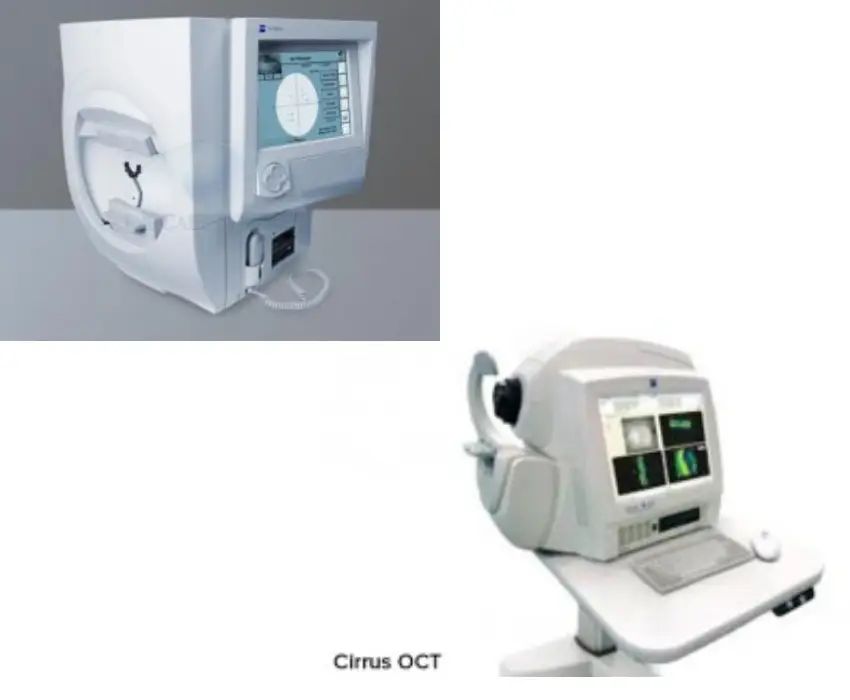
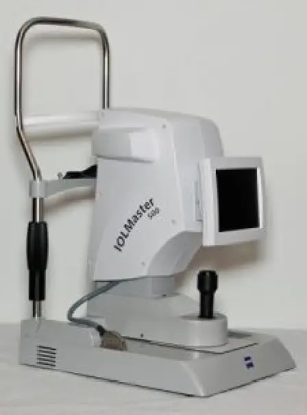
As a comprehensive biometry solution, the IOLMaster® 500 is the preferred method of keratometry for toric IOL calculations1. The IOLMaster 500 now includes the Holladay 2 in its broad menu of recognized formulas for the automatic calculation of the targeted IOL. The IOLMaster 500 is simple, intuitive and fast. It provides precise measurements by providing surgeons the tools they need to optimize outcomes.
It is intended to identify visual field defects for the purposes of screening, monitoring and assisting in the diagnosis and management of occular diseases such as glaucoma and related neurological disorders
Autorefractors are used to measure the degree of refractive error in the eye, and are suited well toward applications such as differentiating corneal from lenticular aberrations, and assessing pre-and-post refractive surgery patients. The patient focuses their vision on a fixation target such as a hot-air balloon floating over land.
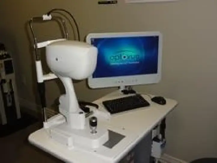
The Optovue OCT provides the newest technology available today for Retina, Optic Nerve, and Cornea imaging. It is instrumental in the diagnosis of Macular Degeneration, Glaucoma, Diabetes, Retinal and Optic Nerve Disorders, Corneal Dystrophies, etc. By performing 26,000 scans per second, this technology provides a 3-Dimensional MRI type image that allows access to the retina and optic nerve that cannot be viewed by other means. This often allows for more accurate diagnoses and better treatment strategies. OCT is used to image the retinal layers. It uses light waves to produce images of the retina, much like sound waves in USG. OCT has opened a new frontier in management of macular diseases.
As a comprehensive biometry solution, the IOLMaster® 500 is the preferred method of keratometry for toric IOL calculations1. The IOLMaster 500 now includes the Holladay 2 in its broad menu of recognized formulas for the automatic calculation of the targeted IOL. The IOLMaster 500 is simple, intuitive and fast. It provides precise measurements by providing surgeons the tools they need to optimize outcomes.
Advanced features such as fundus autofluorescence, easy stereo image handling and innovative assessment of macular pigment optical denstity (MPOD) are combined with intelligent auto functions that enable reproducible and intuitive imaging for every single patient eye.
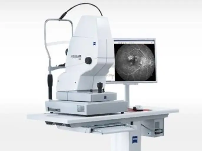
This is a magnified photography of the retina using an injectable dye. It helps in confirming diagnosis, to decide on the mode of treatment and evaluate the treatment given.
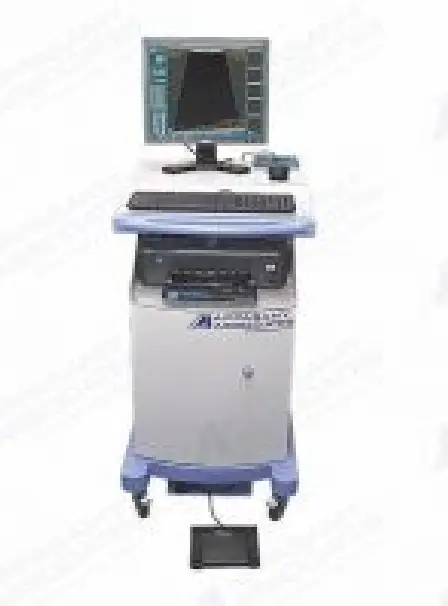
B Probe provide high resolution imaging of the retina and orbit . real _ time video recording play backand editing of B _scan
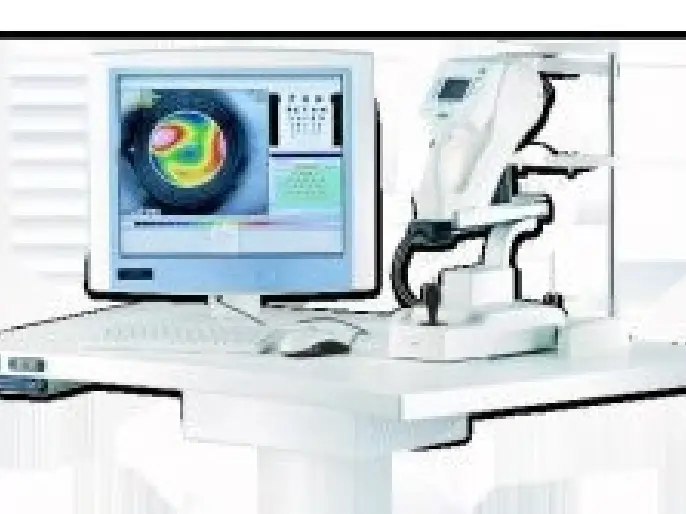
It offers all the possibilites of an innovative topography system. This includes the analysis of corneal wavefront data and thus a particular precise diagnosis in the symptomatic aberrations of the cornea.
B Probe provide high resolution imaging of the retina and orbit . real _ time video recording play backand editing of B _scan
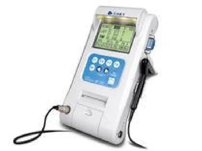
The PalmScan A2000 AScan Biometer is a unique device capable of performing both applanation as well as immersion biometry. It does both quickly and with extreme accuracy.
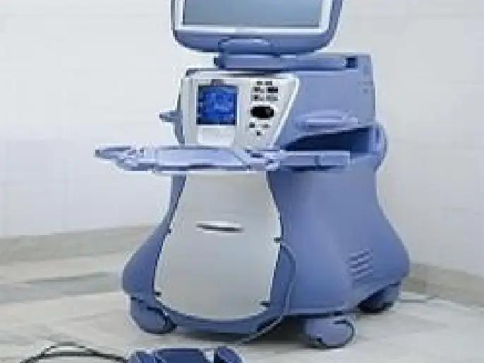
Alcon’s most desirable phaco machine….this Infiniti system is complete and freshly upgraded with 2.05 software, including Ozil and IP Software. We include 1 phaco handpiece, original accessories and the user manual. Our technician also refurbishes the unit to ensure its operating condition. Featuring OZil® Intelligent Phaco software upgrades, the INFINITI® Vision System puts optimized OZil®torsional emulsification at your fingertips. With enhanced fluidic management and surgical control, the INFINITI® Vision System delivers the strategic advantage in customized phaco procedures.
LOCATIONS