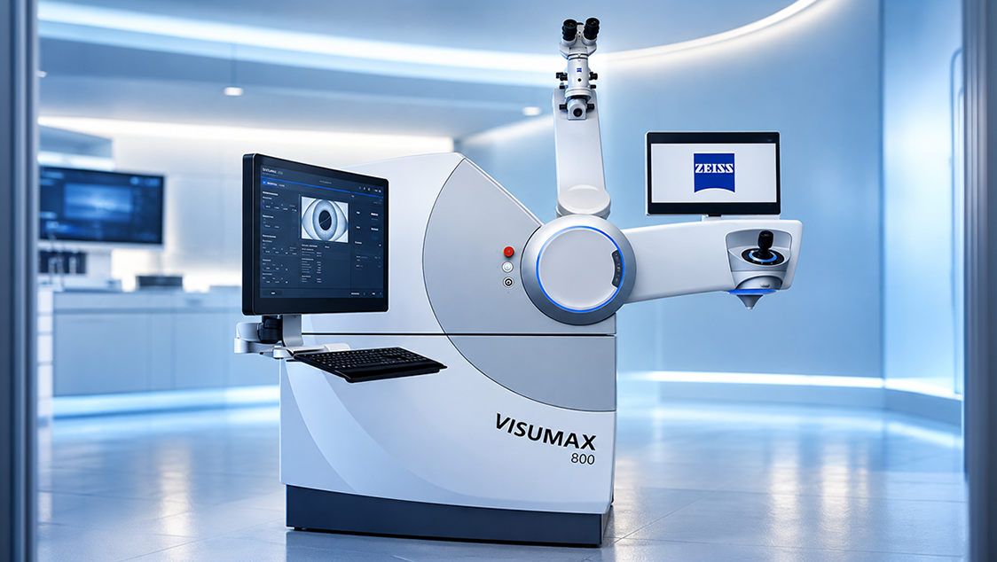
ZEISS VISUMAX 800 is an advanced femtosecond laser system designed for modern refractive procedures such as SMILE Pro and Femto-LASIK. With high-speed laser technology and precise corneal cutting, it supports efficient and controlled vision correction procedures.
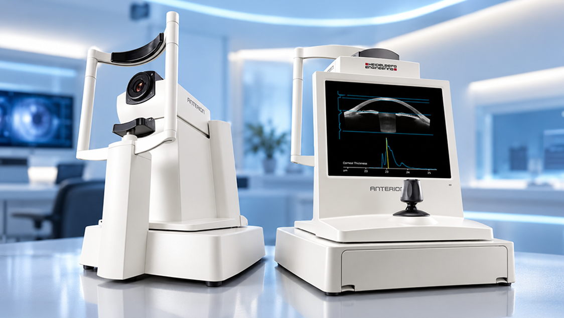
The ANTERION system is an advanced anterior segment imaging platform that uses swept-source OCT technology to deliver high-resolution, cross-sectional images of the eye. It plays a crucial role in evaluating the cornea, anterior chamber, and lens, making it highly valuable for cataract, refractive, and glaucoma assessments.
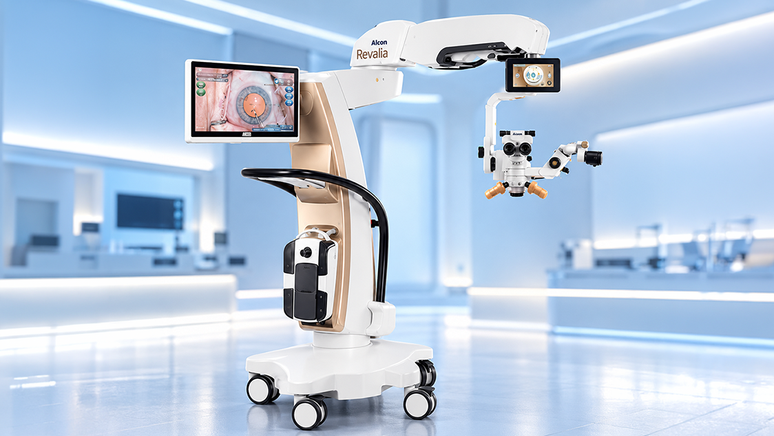
At Abate Eye Hospital, advanced technology plays a vital role in delivering precise and effective eye care. One of the latest innovations available is the ALCON Revalia, designed to enhance surgical accuracy and patient safety.
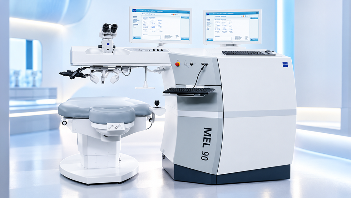
The ZEISS MEL 90 is an advanced excimer laser system designed for modern corneal laser vision correction. It uses precise laser ablation to reshape the cornea and correct refractive errors, supporting procedures such as LASIK and other laser-based treatments for suitable patients.
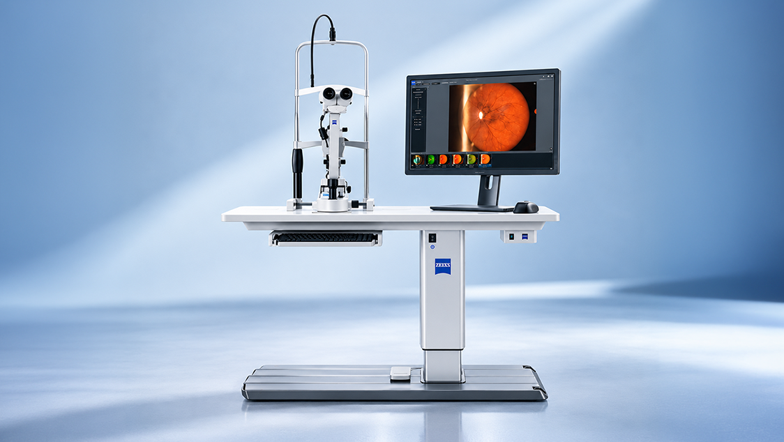
The ZEISS SL 120 Slit Lamp is an advanced ophthalmic examination system designed for detailed examination of the eye. Its high-performance optical system provides clear and precise visualization of the anterior segment, vitreous and fundus, supporting comprehensive eye examinations and accurate clinical assessment.
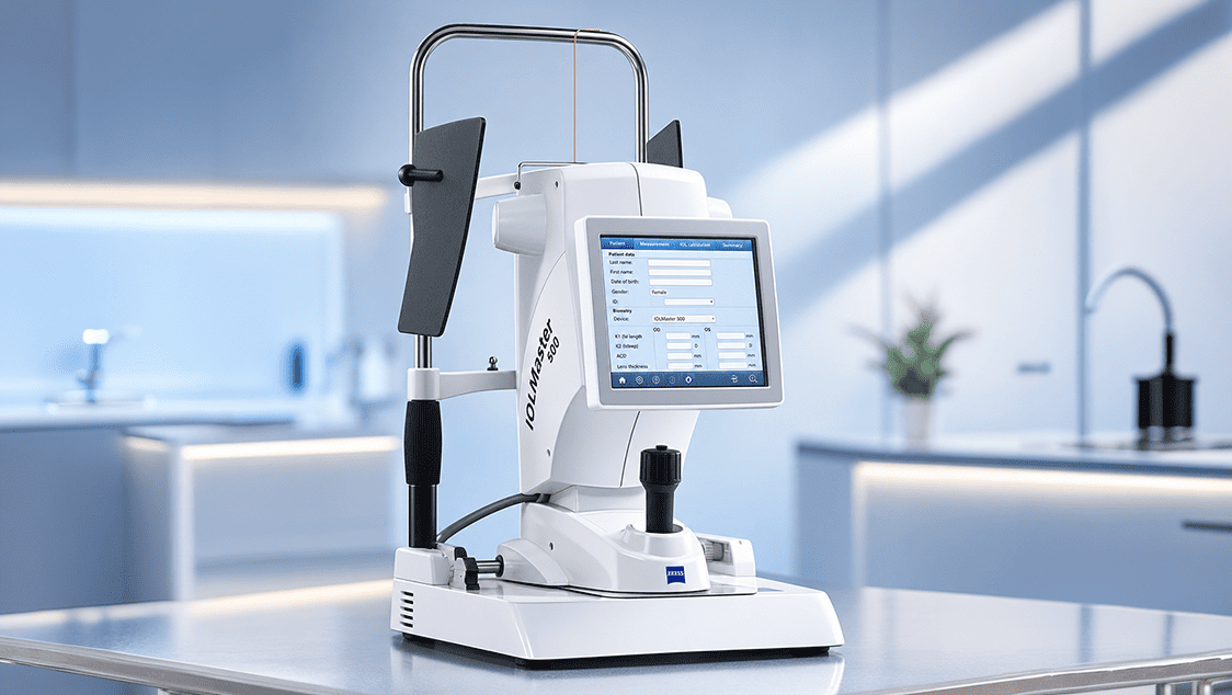
As a comprehensive biometry solution, the IOLMaster® 500 is the preferred method of keratometry for toric IOL calculations. The IOLMaster 500 now includes the Holladay 2 in its broad menu of recognized formulas for the automatic calculation of the targeted IOL. The IOLMaster 500 is simple, intuitive and fast. It provides precise measurements by providing surgeons the tools they need to optimize outcomes.
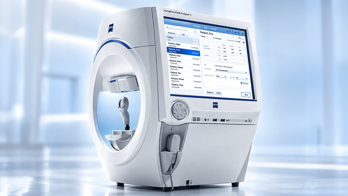
The HUMPHREY HFA FIELD ANALYZER is an advanced visual field testing system used to assess a patient's peripheral and central vision. It helps identify visual field defects and supports the screening, diagnosis, management and monitoring of conditions such as glaucoma and related neurological disorders.
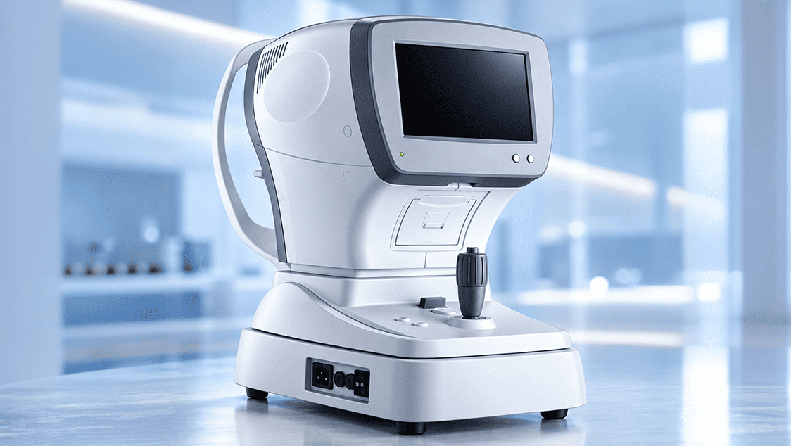
Autorefractors are used to measure the degree of refractive error in the eye, and are suited well toward applications such as differentiating corneal from lenticular aberrations, and assessing pre-and-post refractive surgery patients. The patient focuses their vision on a fixation target such as a hot-air balloon floating over land.
The Optovue OCT provides the newest technology available today for Retina, Optic Nerve, and Cornea imaging. It is instrumental in the diagnosis of Macular Degeneration, Glaucoma, Diabetes, Retinal and Optic Nerve Disorders, Corneal Dystrophies, etc. By performing 26,000 scans per second, this technology provides a 3-Dimensional MRI type image that allows access to the retina and optic nerve that cannot be viewed by other means. This often allows for more accurate diagnoses and better treatment strategies. OCT is used to image the retinal layers. It uses light waves to produce images of the retina, much like sound waves in USG. OCT has opened a new frontier in management of macular diseases.
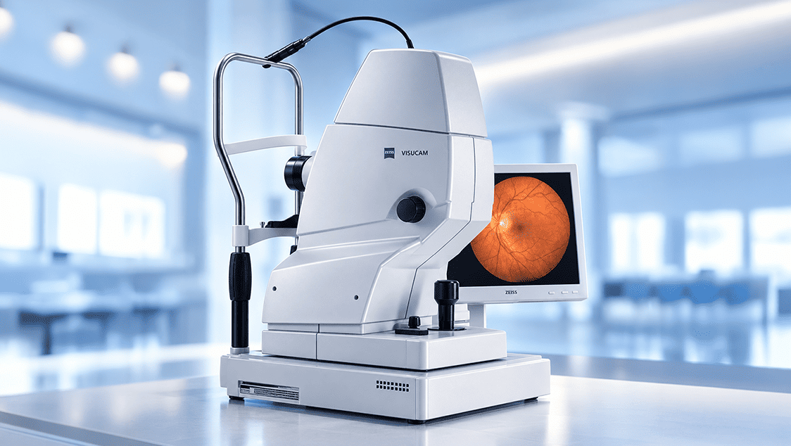
The ZEISS VISUCAM 500 is an advanced digital fundus camera designed to capture detailed images of the retina and the back of the eye. It supports comprehensive retinal examination and documentation, helping ophthalmologists assess retinal health and identify changes associated with various eye conditions.
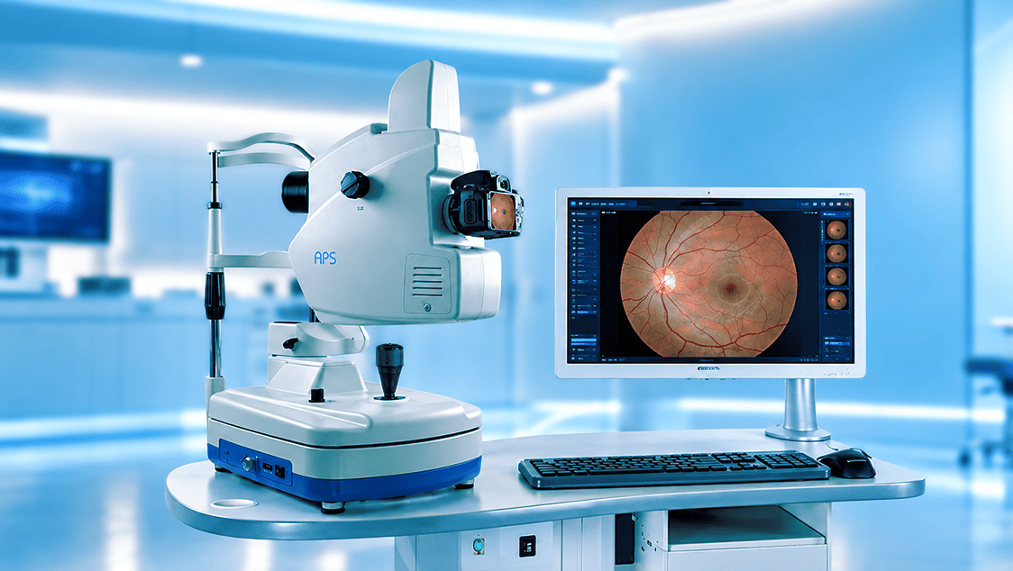
Fundus Fluorescein Angiography (FFA) is an advanced retinal imaging technique used to evaluate the blood circulation within the retina and identify abnormalities in retinal blood vessels. Following the intravenous administration of fluorescein dye, a series of detailed retinal images are captured to help detect leakage, blocked or non-perfused areas and other vascular changes. FFA plays an important role in the diagnosis, treatment planning and monitoring of various retinal conditions.
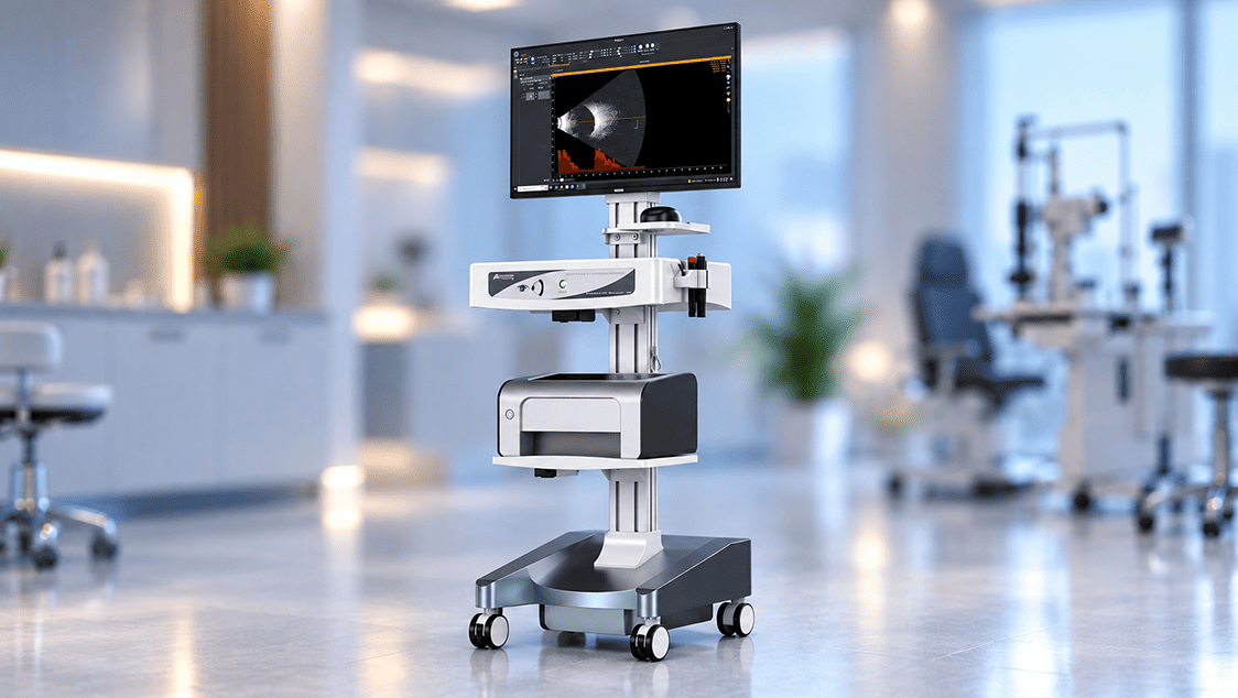
B Probe provide high resolution imaging of the retina and orbit. Real-time video recording, playback and editing of B-scan.
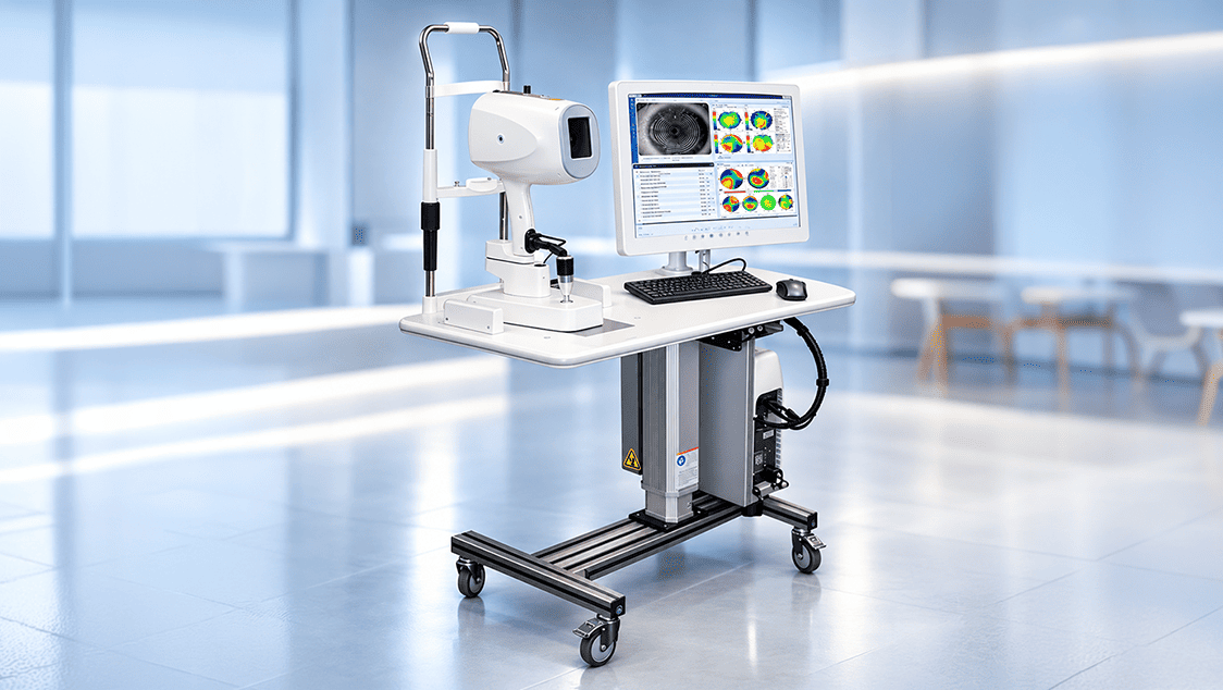
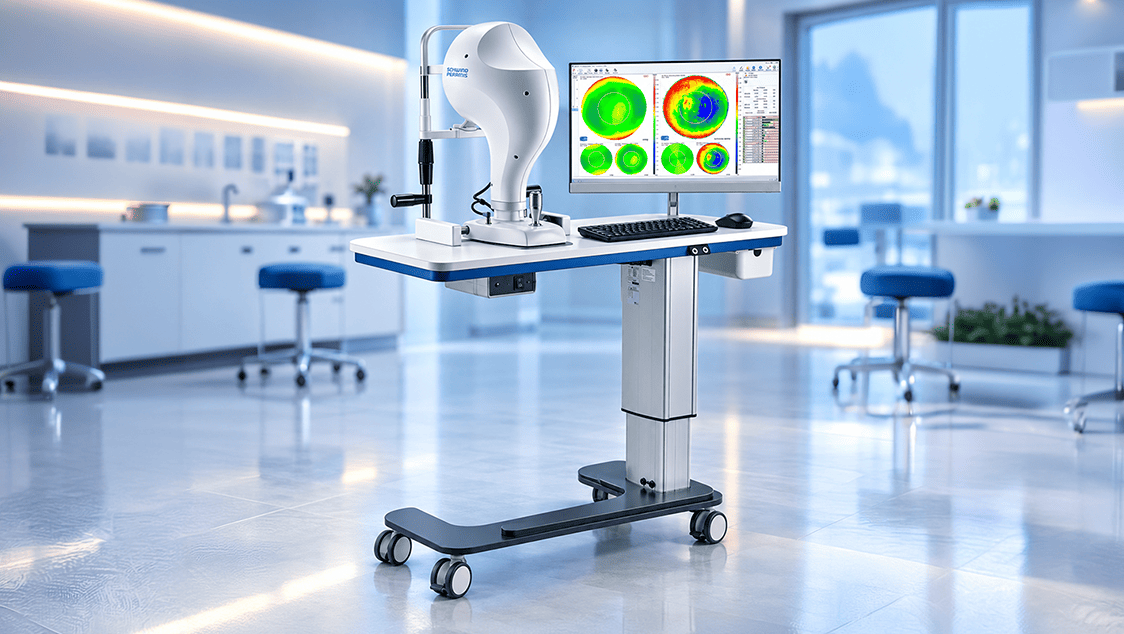
The SCHWIND Corneal Wavefront Analyzer is an advanced diagnostic system designed for detailed assessment of the cornea. It combines corneal topography and wavefront analysis to provide precise information about the shape, surface characteristics and optical properties of the cornea. This detailed evaluation helps ophthalmologists assess corneal conditions and plan personalised refractive and other vision correction procedures.
B Probe provide high resolution imaging of the retina and orbit. Real-time video recording, playback and editing of B-scan.
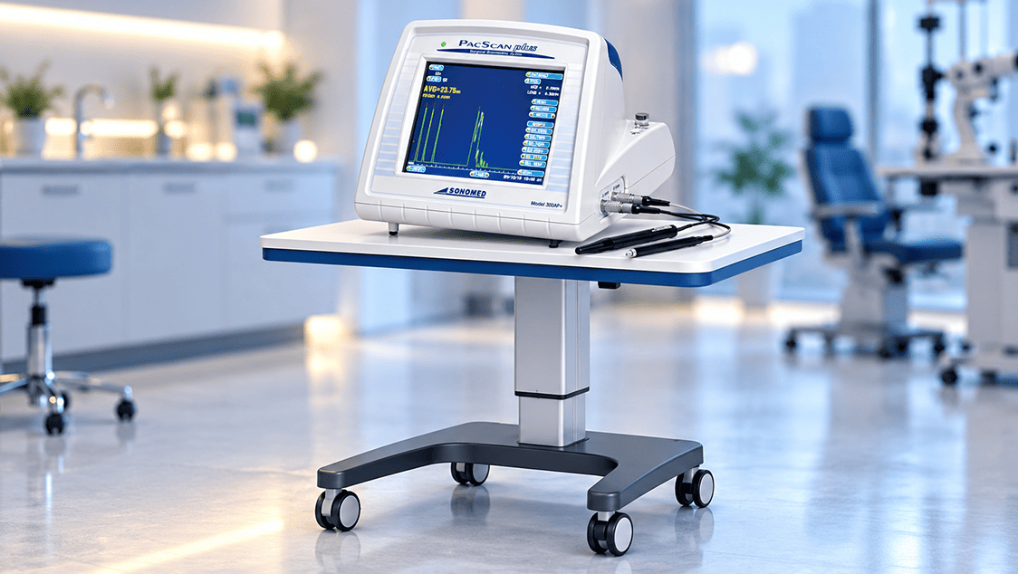
The PalmScan A2000 AScan Biometer is a unique device capable of performing both applanation as well as immersion biometry. It does both quickly and with extreme accuracy.
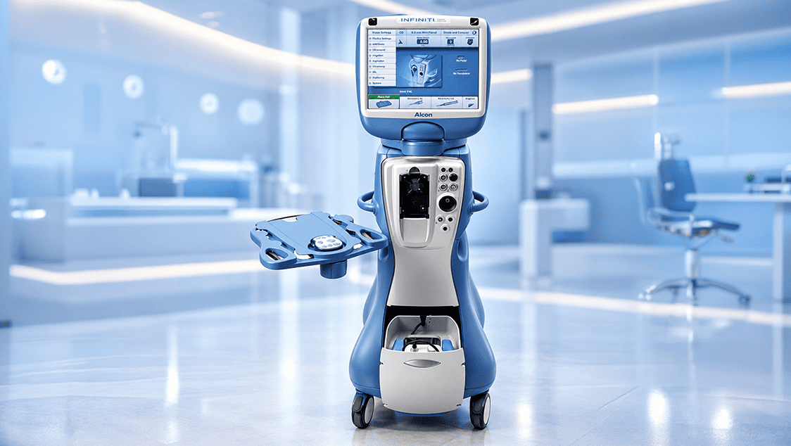
The Alcon INFINITI Vision System is an advanced phacoemulsification platform designed to provide precise and controlled cataract surgery. With OZil® Intelligent Phaco technology, the system uses torsional ultrasound to support efficient lens fragmentation while helping minimise unnecessary movement within the eye. Its advanced fluidics and responsive surgical control allow ophthalmic surgeons to perform cataract procedures with greater precision and consistency.
LOCATIONS